 |
RRUFF Home | UA Mineralogy | Caltech Mineralogy | The IMA Mineral List | Login |

|

|
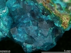
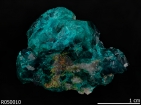
Name: Dioptase RRUFF ID: R050010 Ideal Chemistry: CuSiO3·H2O Locality: Tsumeb mine, Tsumeb, Otavi District, Oshikoto, Namibia Source: Eugene Schlepp Owner: RRUFF Description: Bluish green single crystals Status: The identification of this mineral has been confirmed by X-ray diffraction and chemical analysis |
| Quick search: [ All Dioptase samples (3) ] | ||
| CHEMISTRY | ||||||||||
|---|---|---|---|---|---|---|---|---|---|---|

|
|
|||||||||
| RAMAN SPECTRUM | ||||||||||||||||||||
|---|---|---|---|---|---|---|---|---|---|---|---|---|---|---|---|---|---|---|---|---|
|
||||||||||||||||||||
| BROAD SCAN WITH SPECTRAL ARTIFACTS | ||||||||||||
|---|---|---|---|---|---|---|---|---|---|---|---|---|
|
||||||||||||
| INFRARED SPECTRUM (Attenuated Total Reflectance) | |||||||||||||
|---|---|---|---|---|---|---|---|---|---|---|---|---|---|
|
|||||||||||||
| POWDER DIFFRACTION | ||||||||
|---|---|---|---|---|---|---|---|---|
| RRUFF ID: | R050010.1 | |||||||
| Sample Description: | Powder | |||||||
| Cell Refinement Output: |
a: 14.5759(3)Å b: 14.5759(3)Å c: 7.7821(2)Å alpha: 90.° beta: 90.° gamma: 120.° Volume: 1431.87(6)Å3 Crystal System: hexagonal |
|||||||
|
|
|||||||
| REFERENCES for Dioptase | |
|---|---|
|
American Mineralogist Crystal Structure Database Record: [view record] |
|
|
Anthony J W, Bideaux R A, Bladh K W, and Nichols M C (1990) Handbook of Mineralogy, Mineral Data Publishing, Tucson Arizona, USA, by permission of the Mineralogical Society of America. [view file] |
|
|
Delamétherie J C (1793) De la cristallisation d'une émeraude, Observations sur la Physique, sur l’Histoire Naturelle et sur les Arts, 42, 154-154 [view file] |
|
|
Haüy R J (1797) Dioptase (N.N.), c'est-à-dire, visible au travers, Journal des Mines, 5, 274-275 [view file] |
|
|
Hauy C (1798) Sur la dioptase, Bulletin des Science, par la Société Philomathique, 1798, 101-101 [view file] |
|
|
Vauquelin L N (1798) De la dioptase de Hauy, émeraudine de delamétherie. Essai dur la dioptase, Observations sur la Physique, sur l’Histoire Naturelle et sur les Arts, 46, 301-302 [view file] |
|
|
Belov N V (1957) New silicate structures, Acta Crystallographica, 10, 757-758 |
|
|
Adams D M, Gardner I R (1974) Single-crystal vibrational spectra of beryl and dioptase, Dalton Transactions, 1974, 1502-1505 |
|
|
Belov N V, Maksimov B A, Nozik Y Z, Muradyan L A (1978) The refinement of the crystal structure of dioptase Cu6(Si6O18)(H2O)6 by the X-ray and neutron diffraction methods, Doklady Akademii Nauk SSSR, 239, 842-845 [view file] |
|
|
McKeown D A, Kim C C, Bell M I (1995) Vibrational analysis of dioptase Cu6Si6O18·6(H2O) and its puckered six-member ring, Physics and Chemistry of Minerals, 22, 137-144 |
|
|
Goryainov S V (1996) Dehydration-induced changes in the vibrational states of dioptase, Journal of Structural Chemistry, 37, 58-64 |
|
|
Shannon R D, Shannon R C, Medenbach O, Fischer R X (2002) Refractive index and dispersion of fluorides and oxides, Journal of Physical and Chemical Reference Data, 31, 931-970 [view file] |
|
|
Stebbins F J (2017) Toward the wider application of 29Si NMR spectroscopy to paramagnetic transition metal silicate minerals: copper (II) silicates, American Mineralogist, 102, 2406-2414 |
|
|
Qin F, Wu X, Qin S, Zhang D, Prakapenka V B, Jacobsen S D (2018) Pressure-induced dehydration of dioptase: A single-crystal X-ray diffraction and Raman spectroscopy study, Comptes Rendus Geoscience, In Press, 1-8 [link] |
|
|
|