| RRUFF Home | UA Mineralogy | Caltech Mineralogy | The IMA Mineral List | Login | |
Important Update News
The RRUFF Project is being updated to improve its interface and content. The beta version of the update is accessible to the public at RRUFF.net. New data is only being added to the beta site. Please note that it is in development, and some components are not functional. Existing RRUFF.info links will resolve to the new site after RRUFF.net is officially released.
We are grateful to NASA for the funding of this effort.
)
|
)
|
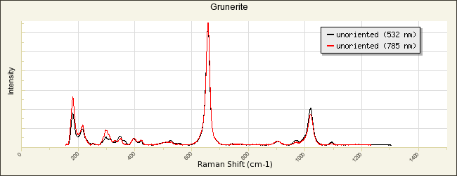
Name: Grunerite RRUFF ID: R070267 Ideal Chemistry: ◻Fe2+2Fe2+5Si8O22(OH)2 Locality: Långfall mine (Saxberg mine), Saxberget ore field, Ludvika, Dalarna, Sweden Source: Michael Scott S101449 [view label] Owner: RRUFF Description: Mass of interlocking light to dark brown striated prismatic crystals associated with sphalerite, garnet and arsenopyrite Status: The identification of this mineral is confirmed by single-crystal X-ray diffraction and chemical analysis. |
| Mineral Group: [ amphibole (107) ] | ||
| Quick search: [ All Grunerite samples (6) ] | ||
| CHEMISTRY | ||||||||||
|---|---|---|---|---|---|---|---|---|---|---|
)
|
|
|||||||||
| RAMAN SPECTRUM | ||||||||||||
|---|---|---|---|---|---|---|---|---|---|---|---|---|
|
||||||||||||
| BROAD SCAN WITH SPECTRAL ARTIFACTS | ||||||||||||
|---|---|---|---|---|---|---|---|---|---|---|---|---|
|
||||||||||||
| POWDER DIFFRACTION | |||||||
|---|---|---|---|---|---|---|---|
| RRUFF ID: | R070267.9 | ||||||
| Sample Description: | Single crystal, powder profile is calculated | ||||||
| Cell Refinement Output: |
a: 9.588(2)Å b: 18.321(5)Å c: 5.335(1)Å alpha: 90° beta: 102.12(1)° gamma: 90° Volume: 916.3(4)Å3 Crystal System: monoclinic |
||||||
|
|
||||||
| REFERENCES for Grunerite | |
|---|---|
|
American Mineralogist Crystal Structure Database Record: [view record] |
|
|
Anthony J W, Bideaux R A, Bladh K W, and Nichols M C (1990) Handbook of Mineralogy, Mineral Data Publishing, Tucson Arizona, USA, by permission of the Mineralogical Society of America. [view file] |
|
|
Kenngott A (1853) VIII. Ordnung: Spathe. IX. Geschlecht: Augit-Spathe. 4. Grunerit, in Das Mohs'sche Mineralsystem Verlag und Druck Wien 62-77 [view file] |
|
|
Winchell A N (1931) Further studies in the amphibole group, American Mineralogist, 16, 250-266 [view file] |
|
|
Ross C S, Kerr P F (1932) The manganese minerals of a vein near Bald Knob, North Carolina, American Mineralogist, 17, 1-18 [view file] |
|
|
Klein C (1964) Cummingtonite-grunerite series: A chemical, optical and x-ray study, American Mineralogist, 49, 963-982 [view file] |
|
|
Finger L W (1969) The crystal structure and cation distribution of a grunerite, Mineralogical Society of America Special Paper, 2, 95-100 [view file] |
|
|
Leake B E (1978) Nomenclature of amphiboles, American Mineralogist, 63, 1023-1052 [view file] |
|
|
Steel E, Wylie A (1981) Mineralogical characteristics of asbestos, 1, in Geology of Asbestos Deposits Edwards Brothers, Inc. Ann Arbor, MI. 93-99 |
|
|
Goldman D S, Rossman G R (1982) The identification of Fe2+ in the M4 site of calcic amphiboles: reply, American Mineralogist, 67, 340-342 [view file] |
|
|
Uchida E (1983) Grunerite from the Shinyama ore deposit, Kamaishi mine, Japan, The Canadian Mineralogist, 21, 517-528 [view file] |
|
|
Hirschmann M, Evans B W, Yang H (1994) Composition and temperature dependence of Fe-Mg ordering in cummingtonite-grunerite as determined by X-ray diffraction, American Mineralogist, 79, 862-877 [view file] |
|
|
Bard D, Yarwood J, Tylee B (1997) Asbestos fibre identification by Raman microspectroscopy, Journal of Raman Spectroscopy, 28, 803-809 [link] |
|
|
Leake B E, Woolley A R, Arps C E S, Birch W D, Gilbert M C, Grice J D, Hawthorne F C, Kato A, Kisch H J, Krivovichev V G, Linthout K, Laird J, Mandarino J A, Maresch W V, Nickel E H, Rock N M S, Schumacher J C, Smith D C, Stephenson N C N, Ungaretti L, Whittaker E J W, Youzhi G (1997) Nomenclature of amphiboles: report of the Subcommittee on Amphiboles of the International Mineralogical Association, Commission on New Minerals and Mineral Names, The Canadian Mineralogist, 35, 219-246 [view file] |
|
|
Huang E P (2002) Raman spectroscopic study of amphiboles, Doctoral Dissertation, 1, 1-138 [view file] |
|
|
Boffa Ballaran T, Carpenter M A (2003) Line broadening and enthalpy: Some empirical calibrations of solid solution behaviour from IR spectra, Phase Transitions, 76, 137-154 |
|
|
Leake B E, Woolley A R, Birch W D, Burke E A J, Ferraris G, Grice J D, Hawthorne F C, Kisch H J, Krivovichev V G, Schumacher J C, Stephenson N C N, Whittaker E J W (2003) Nomenclature of amphiboles: additions and revisions to the International Mineralogical Association’s 1997 recommendations, The Canadian Mineralogist, 41, 1355-1362 [view file] |
|
|
Su S C (2003) A rapid and accurate procedure for the determination of refractive indices of regulated asbestos minerals, American Mineralogist, 88, 1979-1982 [view file] |
|
|
Rinaudo C, Belluso E, Gastaldi D (2004) Assessment of the use of Raman spectroscopy for the determination of amphibole asbestos, Mineralogical Magazine, 68, 455-465 [view file] |
|
|
Roth P (2007) Grunerite, in Minerals first discovered in Switzerland and minerals named after Swiss individuals Kristallografik Verlag Achberg Germany 182-183 |
|
|
Harper M, Lee E G, Doorn S S, Hammond O (2008) Differentiating non-asbestiform amphibole and amphibole asbestos by size characteristics, Journal of Occupational and Environmental Hygiene, 5, 761-770 [view file] |
|
|
Su S C (2008) in How to use the d-spacing/interfacial angle tables to index zone-axis patterns of amphibole asbestos minerals obtained by selected area electron diffraction in transmission electron microscope Asbestos Analysis Consulting Newark, Delaware 1-160 [view file] |
|
|
Apopei A I, Buzgar N (2010) The Raman study of amphiboles, Analele Stiintifice Ale Universitatii, Al. I. Cuza Iasi Geologie, 56, 57-83 [view file] |
|
|
Gunter M E (2010) Defining asbestos: differences between the built and natural environments, Chimia, 64, 747-752 |
|
|
Yong T, Dera P, Zhang D (2019) Single-crystal X-ray diffraction of grunerite up to 25.6 GPa: a new high-pressure clinoamphibole polymorph, Physics and Chemistry of Minerals, 46, 215-227 |
|
|
Germine M, Puffer J H (2020) Analytical transmission electron microscopy of amosite asbestos from South Africa, Archives of Environmental & Occupational Health, 75, 36-44 |
|
|
Tribaudino M, Hovis G L, Almer C, Leaman A (2022) Thermal expansion of minerals in the amphibole supergroup, American Mineralogist, 107, 1302-1312 |
|
|
|