 |
RRUFF Home | UA Mineralogy | Caltech Mineralogy | The IMA Mineral List | Login |

|

|
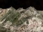
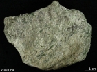
Name: Actinolite RRUFF ID: R040064 Ideal Chemistry: ◻Ca2(Mg4.5-2.5Fe2+0.5-2.5)Si8O22(OH)2 Locality: Ehime Perfecture, Uma Gun, Japan Source: University of Arizona Mineral Museum 5318 [view label] Owner: RRUFF Description: Aggregate of green acicular crystals associated with chlorite Status: The identification of this mineral has been confirmed by X-ray diffraction and chemical analysis |
| Mineral Group: [ amphibole (107) ] | ||
| Quick search: [ All Actinolite samples (11) ] | ||
| CHEMISTRY | ||||||||
|---|---|---|---|---|---|---|---|---|

|
|
|||||||
| RAMAN SPECTRUM | ||||||||||||||||||||
|---|---|---|---|---|---|---|---|---|---|---|---|---|---|---|---|---|---|---|---|---|
|
||||||||||||||||||||
| BROAD SCAN WITH SPECTRAL ARTIFACTS | ||||||||||||
|---|---|---|---|---|---|---|---|---|---|---|---|---|
|
||||||||||||
| INFRARED SPECTRUM (Attenuated Total Reflectance) | |||||||||||||||
|---|---|---|---|---|---|---|---|---|---|---|---|---|---|---|---|
|
|||||||||||||||
| POWDER DIFFRACTION | ||||||||
|---|---|---|---|---|---|---|---|---|
| RRUFF ID: | R040064.1 | |||||||
| Sample Description: | Powder, ~96% actinolite, 4% chlorite | |||||||
| Cell Refinement Output: |
a: 9.8429(3)Å b: 18.0745(7)Å c: 5.2829(2)Å alpha: 90.° beta: 104.714(4)° gamma: 90.° Volume: 909.06(5)Å3 Crystal System: monoclinic |
|||||||
|
|
|||||||
| REFERENCES for Actinolite | |
|---|---|
|
American Mineralogist Crystal Structure Database Record: [view record] |
|
|
Anthony J W, Bideaux R A, Bladh K W, and Nichols M C (1990) Handbook of Mineralogy, Mineral Data Publishing, Tucson Arizona, USA, by permission of the Mineralogical Society of America. [view file] |
|
|
Kirwan R (1794) 16th species: actynolite, in Elements of Mineralogy, 2nd Edition, Volume 1 Elmsly London 167-170 [view file] |
|
|
Winchell A N (1931) Further studies in the amphibole group, American Mineralogist, 16, 250-266 [view file] |
|
|
Hutton C O (1950) Studies of heavy detrital minerals, Bulletin of the Geological Society of America, 61, 635-710 [view file] |
|
|
Klein C (1966) Mineralogy and petrology of the metamorphosed Wabush Iron Formation, Southwestern Labrador, Journal of Petrology, 7, 246-305 |
|
|
Leake B E (1978) Nomenclature of amphiboles, American Mineralogist, 63, 1023-1052 [view file] |
|
|
Goldman D S, Rossman G R (1982) The identification of Fe2+ in the M4 site of calcic amphiboles: reply, American Mineralogist, 67, 340-342 [view file] |
|
|
Spear F S (1982) Phase equilibria of amphibolites from the post pond volcanics, Mt. cube quadrangle, Vermont, Journal of Petrology, 23, 383-426 |
|
|
Dorling M, Zussman J (1985) An investigation of nephrite jade by electron microscopy, Mineralogical Magazine, 49, 31-36 [view file] |
|
|
Arai S, Hirai H (1986) Nickeloan manganoan subcalcic actinolite in a metachert from the Mineoka belt, central Japan, The Canadian Mineralogist, 24, 475-477 [view file] |
|
|
Blount A M (1990) Detection and quantification of asbestos and other trace minerals in powdered industrial-mineral samples, in Process Mineralogy IX The Mineral, Metals & Materials Society, edited by W Petruk, R D Hagni, S Pignolet-Brandom, D M Hausen 557-570 [view file] |
|
|
Leake B E, Woolley A R, Arps C E S, Birch W D, Gilbert M C, Grice J D, Hawthorne F C, Kato A, Kisch H J, Krivovichev V G, Linthout K, Laird J, Mandarino J A, Maresch W V, Nickel E H, Rock N M S, Schumacher J C, Smith D C, Stephenson N C N, Ungaretti L, Whittaker E J W, Youzhi G (1997) Nomenclature of amphiboles: report of the Subcommittee on Amphiboles of the International Mineralogical Association, Commission on New Minerals and Mineral Names, The Canadian Mineralogist, 35, 219-246 [view file] |
|
|
Evans B W, Yang H (1998) Fe-Mg order-disorder in tremolite-actinolite-ferro-actinolte at ambient and high temperature, American Mineralogist, 83, 458-475 [view file] |
|
|
Mikouchi T, Miyamoto M (2000) Micro Raman spectroscopy of amphiboles and pyroxenes in the martian meteorites Zagami and Lewis Cliff 88516, Meteoritics and Planetary Science, 35, 155-159 [view file] |
|
|
Verkouteren J R, Wylie A G (2000) The tremolite-actinolite-ferro–actinolite series: systematic relationships among cell parameters, composition, optical properties, and habit, and evidence of discontinuities, American Mineralogist, 85, 1239-1254 [view file] |
|
|
Huang E P (2002) Raman spectroscopic study of amphiboles, Doctoral Dissertation, 1, 1-138 [view file] |
|
|
Su S C (2003) A rapid and accurate procedure for the determination of refractive indices of regulated asbestos minerals, American Mineralogist, 88, 1979-1982 [view file] |
|
|
Gopal N O, Narasimhulu K V, Rao J L (2004) EPR, optical, infrared and Raman spectral studies of actinolite mineral, Spectrochimica Acta Part A-Molecular and Biomolecular Spectroscopy, 60, 2441-2448 [link] |
|
|
Day H W, Springer R K (2005) The first appearance of actinolite in the prehnite-pumpellyite facies, Sierra Nevada, California, The Canadian Mineralogist, 43, 89-104 [view file] |
|
|
Millette J R, Bandli B R (2005) Asbestos identification using available standard methods, The Microscope, 53, 179-185 |
|
|
Petry R, Mastalerz R, Zahn S, Mayerhöfer T G, Völksch G, Viereck-Götte L, Kreher-Hartmann B, Holz L, Lankers M, Popp J (2006) Asbestos mineral analysis by UV Raman and energy-dispersive X-ray spectroscopy, ChemPhysChem, 7, 414-420 [view file] |
|
|
Harper M, Lee E G, Doorn S S, Hammond O (2008) Differentiating non-asbestiform amphibole and amphibole asbestos by size characteristics, Journal of Occupational and Environmental Hygiene, 5, 761-770 [view file] |
|
|
Su S C (2008) in How to use the d-spacing/interfacial angle tables to index zone-axis patterns of amphibole asbestos minerals obtained by selected area electron diffraction in transmission electron microscope Asbestos Analysis Consulting Newark, Delaware 1-160 [view file] |
|
|
Apopei A I, Buzgar N (2010) The Raman study of amphiboles, Analele Stiintifice Ale Universitatii, Al. I. Cuza Iasi Geologie, 56, 57-83 [view file] |
|
|
Gunter M E (2010) Defining asbestos: differences between the built and natural environments, Chimia, 64, 747-752 |
|
|
Hawthorne F C, Oberti R, Harlow G E, Maresch W V, Martin R F, Schumacher J C, Welch M D (2012) Nomenclature of the amphibole supergroup, American Mineralogist, 97, 2031-2048 [view file] |
|
|
Brown J M, Abramson E H (2016) Elasticity of calcium and calcium-sodium amphiboles, Physics of The Earth and Planetary Interiors, 261, 161-171 |
|
|
Thompson E C, Campbell A J, Liu Z (2016) In-situ infrared spectroscopic studies of hydroxyl in amphiboles at high pressure, American Mineralogist, 101, 706-712 |
|
|
Queffelec A, Fouéré P, Paris C, Stouvenot C, Bellot-Gurlet L (2018) Local production and long-distance procurement of beads and pendants with high mineralogical diversity in an early Saladoid settlement of Guadeloupe (French West Indies), Journal of Archaeological Science: Reports, 21, 275-288 |
|
|
Pieczka A, Stachowicz M, Zelek-Pogudz S, Gołębiowska B, Sęk M, Nejbert K, Kotowski J, Marciniak-Maliszewska B, Szuszkiewicz A, Szełęg E, Stadnicka K M, Woźniak K (2024) Scandian actinolite from Jordanów Śląski, Lower Silesia, Poland: Compositional evolution, crystal structure, and genetic implications, American Mineralogist, 109, 174-183 |
|
|
Su S C in A preliminary characterization of “Libby-type amphiboles” by SAED (Selected Area Electron Diffraction) Batta Labratories, Inc. Newark, Delaware 1-7 [view file] |
|
|
|