| RRUFF Home | UA Mineralogy | Caltech Mineralogy | The IMA Mineral List | Login | |
Important Update News
The RRUFF Project has been migrated to RRUFF.net. Please update your bookmarks immediately, if you have not done so.
The data on this website is already three years out of date, and the entire website will be taken offline before the end of the year.
We are grateful to NASA for the funding of this effort.
)
|
)
|
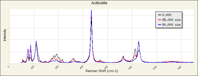
Name: Actinolite RRUFF ID: R060041 Ideal Chemistry: ◻Ca2(Mg4.5-2.5Fe2+0.5-2.5)Si8O22(OH)2 Locality: Prince of Wales Island, Alaska, USA Source: University of Arizona Mineral Museum 4212 [view label] Owner: RRUFF Description: Green acicular crystals forming parallel groups in cylindrical or bladed forms Status: The identification of this mineral has been confirmed by X-ray diffraction and chemical analysis |
| Mineral Group: [ amphibole (107) ] | ||
| Quick search: [ All Actinolite samples (11) ] | ||
| CHEMISTRY | ||||||||||
|---|---|---|---|---|---|---|---|---|---|---|
)
|
|
|||||||||
| RAMAN SPECTRUM | ||||||||||||||||
|---|---|---|---|---|---|---|---|---|---|---|---|---|---|---|---|---|
|
||||||||||||||||
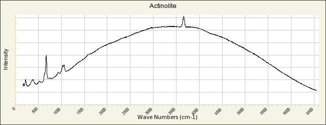
| BROAD SCAN WITH SPECTRAL ARTIFACTS | ||||||||||||
|---|---|---|---|---|---|---|---|---|---|---|---|---|
|
||||||||||||
| INFRARED SPECTRUM (Attenuated Total Reflectance) | |||||||||||||||
|---|---|---|---|---|---|---|---|---|---|---|---|---|---|---|---|
|
|||||||||||||||
| POWDER DIFFRACTION | ||||||||
|---|---|---|---|---|---|---|---|---|
| RRUFF ID: | R060041.1 | |||||||
| Sample Description: | Powder | |||||||
| Cell Refinement Output: |
a: 9.8768(4)Å b: 18.1315(9)Å c: 5.2943(3)Å alpha: 90° beta: 104.747(5)° gamma: 90° Volume: 916.87(6)Å3 Crystal System: monoclinic |
|||||||
|
|
|||||||
| REFERENCES for Actinolite | |
|---|---|
|
American Mineralogist Crystal Structure Database Record: [view record] |
|
|
Anthony J W, Bideaux R A, Bladh K W, and Nichols M C (1990) Handbook of Mineralogy, Mineral Data Publishing, Tucson Arizona, USA, by permission of the Mineralogical Society of America. [view file] |
|
|
Kirwan R (1794) 16th species: actynolite, in Elements of Mineralogy, 2nd Edition, Volume 1 Elmsly London 167-170 [view file] |
|
|
Winchell A N (1931) Further studies in the amphibole group, American Mineralogist, 16, 250-266 [view file] |
|
|
Hutton C O (1950) Studies of heavy detrital minerals, Bulletin of the Geological Society of America, 61, 635-710 [view file] |
|
|
Klein C (1966) Mineralogy and petrology of the metamorphosed Wabush Iron Formation, Southwestern Labrador, Journal of Petrology, 7, 246-305 |
|
|
Leake B E (1978) Nomenclature of amphiboles, American Mineralogist, 63, 1023-1052 [view file] |
|
|
Goldman D S, Rossman G R (1982) The identification of Fe2+ in the M4 site of calcic amphiboles: reply, American Mineralogist, 67, 340-342 [view file] |
|
|
Spear F S (1982) Phase equilibria of amphibolites from the post pond volcanics, Mt. cube quadrangle, Vermont, Journal of Petrology, 23, 383-426 |
|
|
Dorling M, Zussman J (1985) An investigation of nephrite jade by electron microscopy, Mineralogical Magazine, 49, 31-36 [view file] |
|
|
Arai S, Hirai H (1986) Nickeloan manganoan subcalcic actinolite in a metachert from the Mineoka belt, central Japan, The Canadian Mineralogist, 24, 475-477 [view file] |
|
|
Blount A M (1990) Detection and quantification of asbestos and other trace minerals in powdered industrial-mineral samples, in Process Mineralogy IX The Mineral, Metals & Materials Society, edited by W Petruk, R D Hagni, S Pignolet-Brandom, D M Hausen 557-570 [view file] |
|
|
Leake B E, Woolley A R, Arps C E S, Birch W D, Gilbert M C, Grice J D, Hawthorne F C, Kato A, Kisch H J, Krivovichev V G, Linthout K, Laird J, Mandarino J A, Maresch W V, Nickel E H, Rock N M S, Schumacher J C, Smith D C, Stephenson N C N, Ungaretti L, Whittaker E J W, Youzhi G (1997) Nomenclature of amphiboles: report of the Subcommittee on Amphiboles of the International Mineralogical Association, Commission on New Minerals and Mineral Names, The Canadian Mineralogist, 35, 219-246 [view file] |
|
|
Evans B W, Yang H (1998) Fe-Mg order-disorder in tremolite-actinolite-ferro-actinolte at ambient and high temperature, American Mineralogist, 83, 458-475 [view file] |
|
|
Mikouchi T, Miyamoto M (2000) Micro Raman spectroscopy of amphiboles and pyroxenes in the martian meteorites Zagami and Lewis Cliff 88516, Meteoritics and Planetary Science, 35, 155-159 [view file] |
|
|
Verkouteren J R, Wylie A G (2000) The tremolite-actinolite-ferro–actinolite series: systematic relationships among cell parameters, composition, optical properties, and habit, and evidence of discontinuities, American Mineralogist, 85, 1239-1254 [view file] |
|
|
Huang E P (2002) Raman spectroscopic study of amphiboles, Doctoral Dissertation, 1, 1-138 [view file] |
|
|
Su S C (2003) A rapid and accurate procedure for the determination of refractive indices of regulated asbestos minerals, American Mineralogist, 88, 1979-1982 [view file] |
|
|
Gopal N O, Narasimhulu K V, Rao J L (2004) EPR, optical, infrared and Raman spectral studies of actinolite mineral, Spectrochimica Acta Part A-Molecular and Biomolecular Spectroscopy, 60, 2441-2448 [link] |
|
|
Day H W, Springer R K (2005) The first appearance of actinolite in the prehnite-pumpellyite facies, Sierra Nevada, California, The Canadian Mineralogist, 43, 89-104 [view file] |
|
|
Millette J R, Bandli B R (2005) Asbestos identification using available standard methods, The Microscope, 53, 179-185 |
|
|
Petry R, Mastalerz R, Zahn S, Mayerhöfer T G, Völksch G, Viereck-Götte L, Kreher-Hartmann B, Holz L, Lankers M, Popp J (2006) Asbestos mineral analysis by UV Raman and energy-dispersive X-ray spectroscopy, ChemPhysChem, 7, 414-420 [view file] |
|
|
Harper M, Lee E G, Doorn S S, Hammond O (2008) Differentiating non-asbestiform amphibole and amphibole asbestos by size characteristics, Journal of Occupational and Environmental Hygiene, 5, 761-770 [view file] |
|
|
Su S C (2008) in How to use the d-spacing/interfacial angle tables to index zone-axis patterns of amphibole asbestos minerals obtained by selected area electron diffraction in transmission electron microscope Asbestos Analysis Consulting Newark, Delaware 1-160 [view file] |
|
|
Apopei A I, Buzgar N (2010) The Raman study of amphiboles, Analele Stiintifice Ale Universitatii, Al. I. Cuza Iasi Geologie, 56, 57-83 [view file] |
|
|
Gunter M E (2010) Defining asbestos: differences between the built and natural environments, Chimia, 64, 747-752 |
|
|
Hawthorne F C, Oberti R, Harlow G E, Maresch W V, Martin R F, Schumacher J C, Welch M D (2012) Nomenclature of the amphibole supergroup, American Mineralogist, 97, 2031-2048 [view file] |
|
|
Brown J M, Abramson E H (2016) Elasticity of calcium and calcium-sodium amphiboles, Physics of The Earth and Planetary Interiors, 261, 161-171 |
|
|
Thompson E C, Campbell A J, Liu Z (2016) In-situ infrared spectroscopic studies of hydroxyl in amphiboles at high pressure, American Mineralogist, 101, 706-712 |
|
|
Queffelec A, Fouéré P, Paris C, Stouvenot C, Bellot-Gurlet L (2018) Local production and long-distance procurement of beads and pendants with high mineralogical diversity in an early Saladoid settlement of Guadeloupe (French West Indies), Journal of Archaeological Science: Reports, 21, 275-288 |
|
|
Pieczka A, Stachowicz M, Zelek-Pogudz S, Gołębiowska B, Sęk M, Nejbert K, Kotowski J, Marciniak-Maliszewska B, Szuszkiewicz A, Szełęg E, Stadnicka K M, Woźniak K (2024) Scandian actinolite from Jordanów Śląski, Lower Silesia, Poland: Compositional evolution, crystal structure, and genetic implications, American Mineralogist, 109, 174-183 |
|
|
Su S C in A preliminary characterization of “Libby-type amphiboles” by SAED (Selected Area Electron Diffraction) Batta Labratories, Inc. Newark, Delaware 1-7 [view file] |
|
|
|